Getting Started
The main purpose of this page is to show you how to quickly start segmenting new MRIs using HSF.
Installation
This is a quick getting-started guide. For in-depth details, please check the page dedicated to installation details.
Setting up a new environment
We encourage you to use the conda environment manager
to use HSF.
To create a new environment, run the following command:
conda create -n hsf python=3.9
Then, activate the new environment:
source activate hsf
Basic installation
Once the environment is set up, you can install HSF using the following command:
pip install hsf["cpu"]
Hardware acceleration
By doing so, HSF will be limited to your CPU. If you want to use a different hardware acceleration, please refer to the page dedicated to installation details.
You can now check the installation and default parameters of HSF using
hsf -h
Say hello to your new segmentation!
Now, let's say you want to segment a database of MRIs, comprising CoroT2 images of anisotropic resolution 0.3x1.0x0.3.
We do not impose any specific constraints on the database structure, but please take in consideration that file names should be unambiguous and easily recognizable through a glob.
Here is an example of a database structure:
Database
│   README.md
│   CHANGELOG.txt
│
└───sub-00C
│   │   sub-00C_tse.nii.gz
│   │   sub-00C_t1w.nii.gz
│
└───sub-01C
    │   sub-01C_tse.nii.gz
    │   sub-01C_t1w.nii.gz
To segment the whole database, you can just run the following command:
hsf files.path="~/Database" files.pattern="**/*tse.nii.gz" roiloc.margin=[16,4,16]
And that's it! 
Now, do you want to segment the T1w MRIs? Nothing simpler:
hsf files.path="~/Database" files.pattern="**/*t1w.nii.gz" roiloc.margin=[8,8,8] roiloc.contrast="t1"
files.path and files.pattern are the only mandatory parameters. They are used to
determine the list of files to segment.
The parameter roiloc.margin is not mandatory, but one of the most important parameters
of HSF. It defines the size of the margin around each hippocampi.
A too small margin will likely result in incorrect crops of the hippocampi, thus leading to a truncated segmentation. It will also decrease the accuracy of the segmentation as it is removing context in the image. A large margin however, is likely to increase the accuracy of the segmentation, but it will greatly increase the computational requirements.
Keep the box centered around the hippocampus, but not too close to it.
Here is a good example:
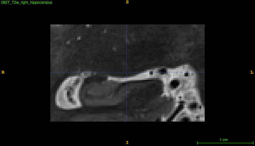
If you want to check the resulting boxes, HSF creates a folder for each subject called
hsf_outputs, where you'll find boxes for each of the segmented hippocampi.
For more details, please check the dedicated page.